|
|
|
The mission of Laboratory of Optical Studies for Advanced Materials (LOSAM) is to develop and characterize frontier materials and devices with advanced optical techniques. To pursue this goal, the laboratory is equipped with (1) high-power femtosecond laser facility, (2) widely tunable picosecond coherent light sources; and (3) Yb-doped fiber laser pumped femtosecond laser with pulse shaping apparatus, etc. To probe into material properties and structures, we have developed several advanced spectroscopies and microscopies including infrared-visible sum-frequency vibrational spectroscopy (SFVS), scanning near field optical microscopy (SNOM), single-molecular detection and imaging apparatus, and 2D coherent spectroscopy (2D CS). |
 Single-Molecule optical Microscopy (SMoM)The single-molecule probing method has become a valuable tool in single-molecular biophysics. If an optical process is employed, the resulting technique can possess both single-molecule detection (SMD) sensitivity and minimum invasiveness. Optical probes have therefore become the most attractive choice in single-molecule biophysics. By using photo-activatable fluorophores, one can modulate the fluorescence emission profile of individual fluorophores in time such that only an optically resolvable subset of fluorophores are activated at any moment, allowing their localization with high accuracy. It has been realized that photo-activatable fluorophores are becoming the bottleneck of the wide-field sub-diffraction imaging technique. Therefore, to facilitate the development of new robust molecular photo-switches, we are investigating the mechanism of photo-activation and exploring the potential of sub-diffraction imaging/localization of a variety of biomolecules. Profound issues such as the interaction between cell-penetrating peptides and cell membrane, mechanisms of protein-drug interaction in a live cell are to be studied in this project. More... 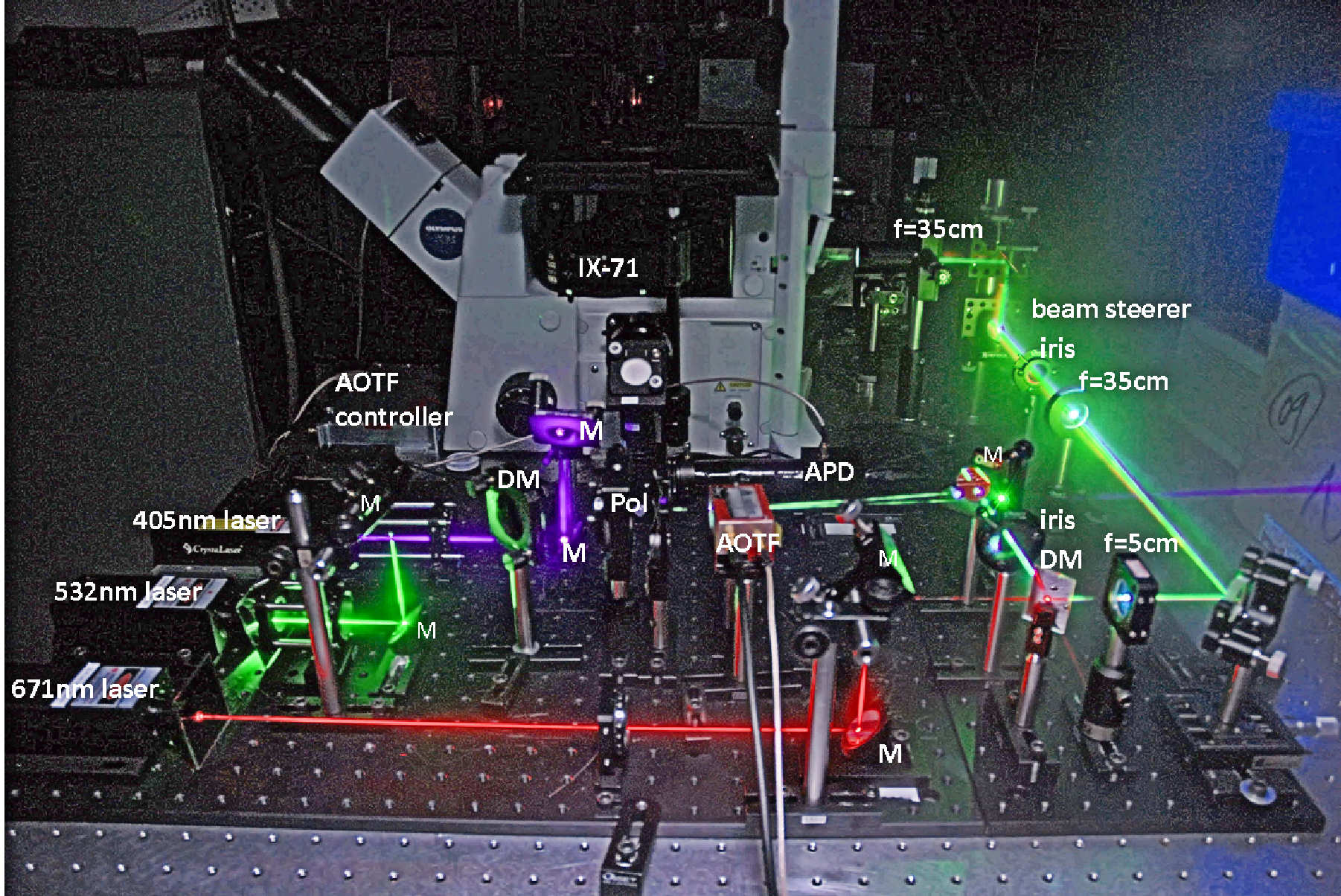
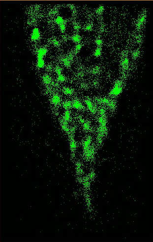
FIG. 1 (Left) Multiwavelength optical microscope employed for single-molecular detection and imaging. (Right) Superresolution image of the plasma membrane of a live He-La cell labeled with DII dyes. |
Femtosecond 2D Coherent SpectroscopyTracking correlated intramolecular and intermolecular motions are crucial for understanding complex molecular systems. We are able to use the time-resolved asynchronous scan FTIR technique to probe the switching dynamics of pure ferroelectric liquid crystal (FLC) and FLC mixtures. The study revealed the detailed correlated motion of submolecular fragments in a FLC film and can distinguish specific functional groups attached to different positions on a FLC molecule. 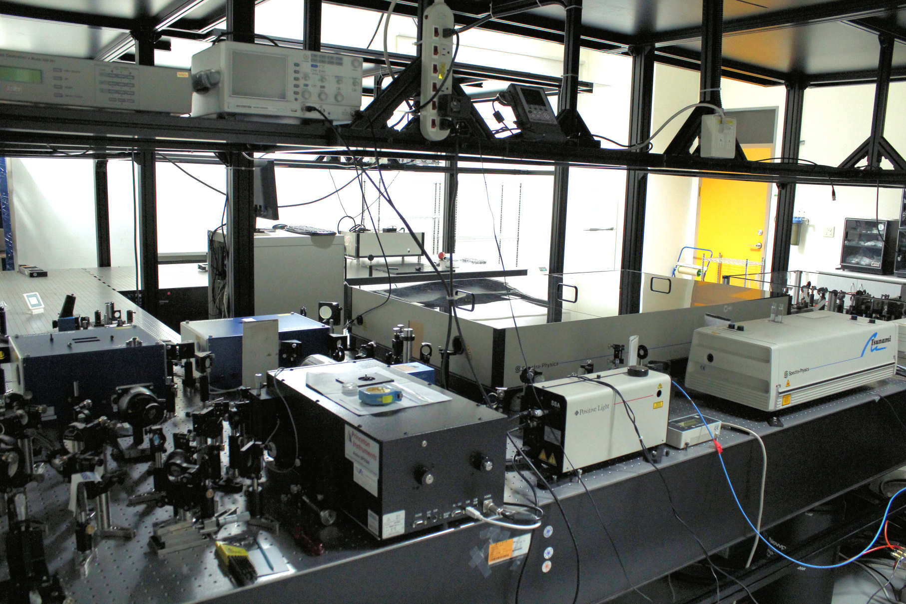
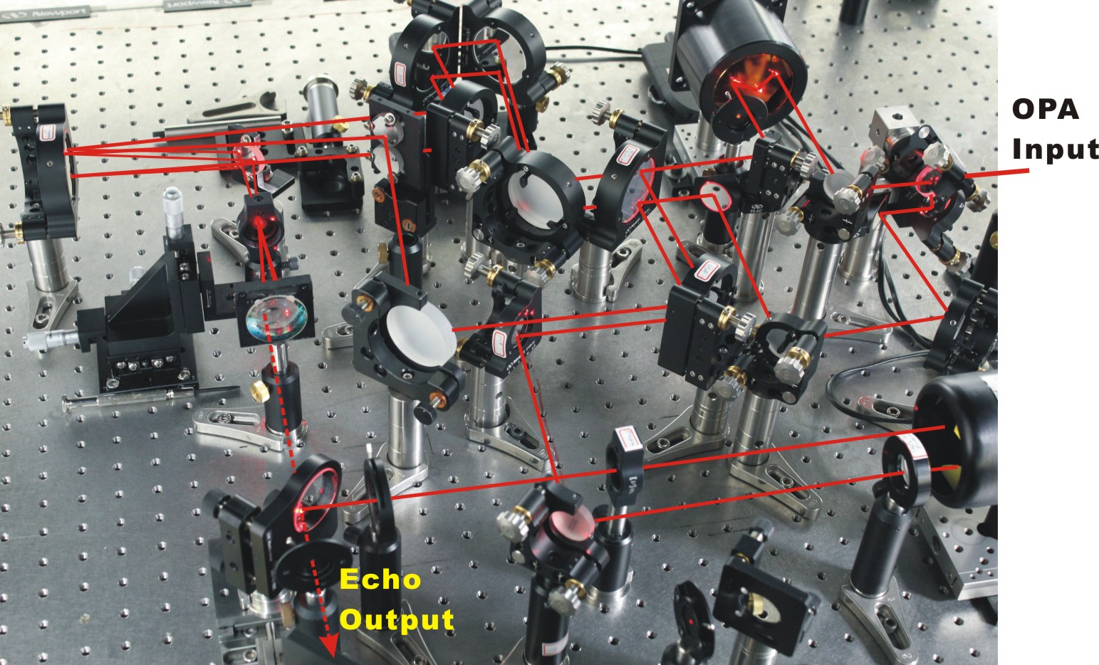
FIG. 2. High-power femtosecond laser facility (left) and the 2D correlation spectroscopy installed in the labratory for investigating dynamics of semiconductor nanostructures and biomolecules. The important feature of the time-resolved asynchronous scan FTIR technique is that the achievable temporal resolution is limited only by the IR pulsewidth used. By using the state-of-the-art techniques of laser and nonlinear optics, femtosecond IR pulses with wide bandwidth and short temporal duration can be generated for the time-resolved 2D IR applications. The time-resolved 2D IR spectroscopy can probe the couplings between vibrational modes, which are related to their relative orientations, and hence allows for extraction of structural information. Vibration-vibration coupling is sensitive to structure in a manner analogous to measurements of spin-spin coupling in 2-D NMR. The most promising advantage of 2D-IR spectroscopy is its intrinsic high temporal resolution (<1 ps), which freezes in all motions of the system. This allows one to resolve rapidly exchanging conformations of an equilibrium ensemble of systems. One can monitor how a non-equilibrium ensemble evolves in real time with subpicosecond time resolution. Undoubtedly, the progress made in optical pulse-shaping techniques and new interferometric measuring techniques in recent years will have an enormous impact on these very promising fields. The 2D Visible/IR coherent spectroscopy installed in our laboratory is based on fs Ti:saphire laser (CW mode-locking and then regeneratively amplified to 2 mJ at 1kHz with 50 fs pulsewidth). Wavelength extension is achieved with nonlinear optical parametric generation and difference-frequency generation scheme. The following interesting studies are undertaking to demonstrate the cutting-edge techniques and to advance our knowledge of the structural dynamics of semiconductor nanostructures and biological molecular systems: I. The exciton coupling dynamics of energy harnessing materials; and II. The dynamics mediated by electron-phonon coupling in InAs quantum dots embedded in an asymmetric quantum well structure. More... |
Sum-Frequency Vibrational Spectroscopy (SFVS)Infrared-visible SFVS and second-harmonic generation (SHG) have been demonstrated to be effective tools to probe molecular structures and orientations on a variety of surfaces and interfaces. They have the advantage of being highly surface-specific and sensitive. The surface specificity arises because under the electric-dipole approximation, these processes are forbidden in media with inversion symmetry, but allowed at a surface or interface where such symmetry is broken. For more detailed applications of SFVS on polymeric surface, please press here. In nearly all electro-optic applications of liquid crystals (LC), it is necessary to uniformly align the liquid crystal molecules to along a certain direction. Aligned polar structure of liquid crystal can be formed in a surface-stabilized ferroelectric liquid crystal (SSFLC) film. Polar structure of SSFLC can yield a nonvanishing dipole moment per unit volume Ps, which is switchable with an electric field E via an interactive energy of -Ps.E. The resulting switching speed of SSFLC is much higher than that with nematic LC. To yield a clearer picture of the physical mechanism of LC alignment by polymer surface, an investigation of LC aligned structure on a number of different polymeric films is highly desirable. The latter information is often difficult to be obtained with SHG measurement alone. The following apparatus based on a highpower picosecond laser system and nonlinear optical techniques had been installed in my laboratory to allow us pursue the goal. More... 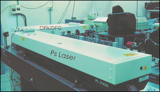
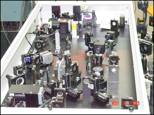
FIG. 3. (Left) Picosecond laser and optical parametric amplification (OPA)/difference frequency generation (DFG) systems used for SFVS. (Right) Detailed setup of the OPA/DFG for the generation of tunable infrared pulses from 2.5 um to 10 um. 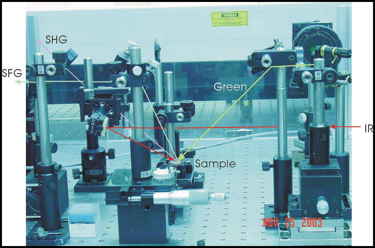
FIG. 4. Optical setup of SFVS for studying molecular structures and orientations of varying polymer surfaces for aligning liquid crystal molecules. |
Heterodyne SNOMThe key to successful developments of functional photonic devices lies in the fabrication and characterization. Valuable diagnostic tools not only improve our knowledge of photonic devices but also help us to establish the design rules. In view of this impact, during the past years we had initiated an effort to develop a diagnostic platform for nanophotonic devices. 
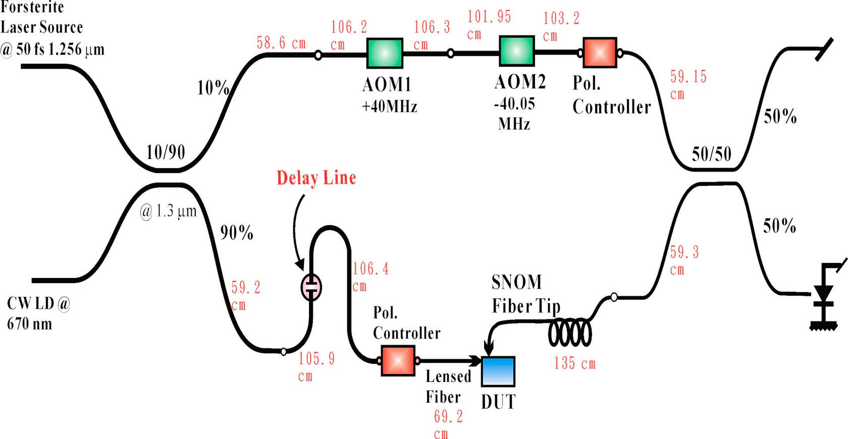
FIG. 5. (Left) Photograph showing the real setup for an all-fiberized hetrodyne scanning near field optical microscope installed in my laboratory. The entire system is located in an enclosure to prevent air turbalance and providing acoustic shielding. (Right) Schematic diagram showing the all-fiberized heterodyne interferometer. The diagnostic platform includes a heterodyne interferometer to yield unique advantages of shot-noise limited detection sensitivity and infinite phase tracking capability. 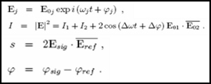
Here the signal intensity Is ~1x10-12 W ~1x10+7 photons/sec is below the noise floor of photodiode detectors. But by interfering this signal with Iref~1x10⁻⁴ W , the signal at the detector can be boosted to Is~1x10⁻⁸ W , which is well within the detection limit of a typical photo-detector. In order to reveal the full-field characteristics of photonic devices, we combine the heterodyne interferometer with a scanning near field optical microscopy (SNOM) for the development of photonic devices with engineered subwavelength structures. Furthermore to couple light field efficiently into a photonic device, an efficient light coupling scheme with a lensed optical fiber into a photonic device in a SNOM setup is used. |
Femtosecond Pulse ShaperThe purpose of femtosecond coherent control study is not only to control the evolution of a complex system but also to deduce the detailed dynamic mechanism from the optimal laser field used. Recently, we have developed an adaptive femtosecond pulse shaper (see Fig. 4: Left) for rapid quantum control. With the technique the desirable optical field characteristics can be verified and be used to selectively excite one type of probe molecule while leave the others in their ground state. The coherent control methodology also offers an additional degree of freedom to distinguish coherent and incoherent nonlinear optical processes. We combined a scanning optical microscope (see Fig. 4: Right) with a femtosecond pulse shaper to achieve selective mapping of the distribution of specific species with coherent control methodology. 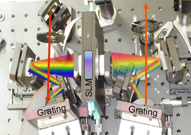
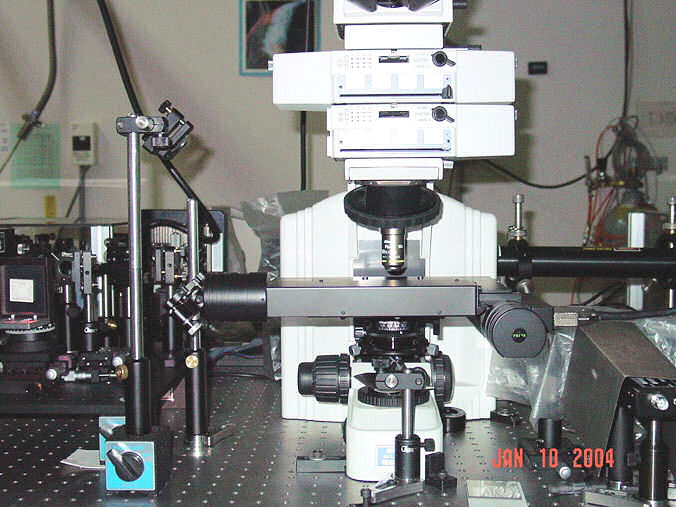
FIG. 6. (Left) Femtosecond pulse shaper, (Right) Scanning optical microscope employed for molecular specific mapping with ultrafast coherent control methodology. To analyze extremely weak light-material interaction in real time, we also developed for the first time a single-shot OPA-FROG, which can be used to completely characterize a coherent optical field as weak as several atto joules. Complete-field characterization of advanced photonic materials and devices at the nanometer scales is also undertaking by combining scanning near-field optical microscopy (SNOM) with heterodyne interferometry. |